Types Of Hemangioma Radiology
There are three main types of hemangioma. This article aims to be a generic discussion of the condition for detailed and more specific imaging features please refer to subarticles.
 Vertebral Hemangioma Radiology Reference Article Radiopaedia Org
A hemangioma is a common vascular birthmark made of extra blood vessels in the skin.

Types of hemangioma radiology. In contrast a rare type of vascular tumor intramuscular capillary-type hemangioma usually presents beyond the period of infancy with nonspecific symptoms and no evidence of involution. There are many types of hemangiomas and they can occur throughout the body including in skin muscle bone and internal organs. Hemangiomas with fluid-fluid levels.
Klippel-Trenaunay-Weber disease Osler-Rendu-Weber disease and von HippelLindau disease are all associated with hemangiomas 3. Less frequent types are large heterogeneous hemangiomas. They are slow-growing and most are not symptomatic.
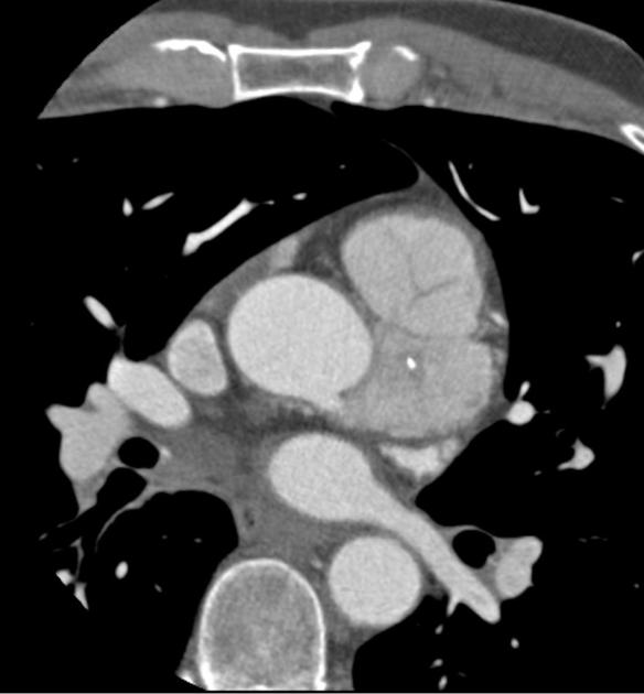
Hemangiomas present a diagnostic challenge because they can be mistaken for hyper-vascular malignancies of the liver and can coexist with and occasionally mimic other benign and malignant hepatic lesions including focal nodular hyperplasia hepatic adenoma hepatic cysts hemangio-endothelioma hepatic metastasis and primary hepatocellular carcinoma 1 2 3. Hepatic cavernous venous malformation hepatic hemangioma atypical hepatic venous malformation atypical hepatic hemangioma giant hepatic venous malformation giant hepatic hemangioma flash filling hepatic venous malformation flash filling hepatic hemangioma splenic cavernous venous malformation splenic hemangioma. The differential diagnosis of spinal epidural hemangiomas included herniated disks synovial cysts granulomatous infections neurogenic tumors lymphomas meningiomas angiolipoma pure epidural hematoma and epidural extramedullary hematopoiesis depending on the MR types 5 8 11 14 19 On-line Table 2.
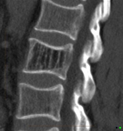
T1 T2 and STIR MRI images of a vertebral hemangioma A vertebral hemangioma VH is a vascular lesion within a vertebral body. In some cases specifically capillary types lytic erosion into the epidural space can occur however rare 2. On unenhanced CT it may appear as an ill-defined mass of similar attenuation to muscle.
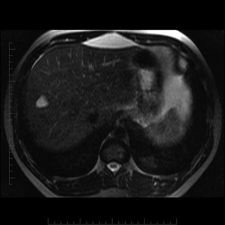
A liver hemangioma is made up of a tangle of blood vessels. 1 Axial view of focal hepatic hemangioma demonstra-. Small lesions may be occult on plain film while large lesions may show evidence of a focal soft tissue swelling - associated.
Other terms for a liver hemangioma are hepatic hemangioma and cavernous hemangioma. Superficial on the surface of the skin deep under the skin and mixed. 1 2 However some lesions are known as mixed-type haemangiomas because they have an admixture of different types of vessels.
Based on this analysis we speculated that IHH comprised 3 subtypes. Capillary cavernous venous and arteriovenous AV haemangiomas. It has two main histopathological types cavernous involves relatively large vessels and capillary involves small capillaries angiomas 11.
In the borderline category kaposiform hemangioendothelioma is a childhood tumor that may be associated with thrombocytopenia and consumptive coagulopathy whereas Kaposi sarcoma is most commonly encountered in the immunocompromised adult. A frequent type of atypical hepatic hemangioma is a lesion with an echoic border at ultrasonography. The various retinal vascular tumors are benign and each has distinct funduscopic and imaging features.
Haemangiomas are pathologically classified into four types based on the predominant type of vascular channel identified within the lesion. Infantile hemangiomas are benign vascular neoplasms that are the most common head and neck tumors of infancy. Infantile hemangiomas demonstrate a pattern of proliferative growth in infancy followed by a slow phase of involution.
CT may also show the. Cystic or multilocular hemangiomas. A liver hemangioma he-man-jee-O-muh is a noncancerous benign mass in the liver.
Progressive peripheral enhancement with more. Most cases of liver hemangiomas are discovered during a test or procedure for some other condition. They can occur virtually anywhere but the majority are found in the head and neck regions.
Features of typical lesions include. Vertebral hemangiomas are a common etiology estimated to be found in 10-12 of humans at autopsy. Several types of benign and malignant tumors can arise in the retina originating from neural retinoblastoma vascular hemangiomahemangioblastoma and glial astrocytic hamartoma and acquired astrocytoma elements.
Focal multifocal and diffuse with each category demon-strating distinctive imaging pathologic and physiologic Fig. They often develop on the face and neck and can vary greatly in color shape and size. Most hemangiomas occur on the surface of the skin or just beneath it.
Commonly these are benign lesions that are found incidentally during radiology studies for other indications. At ultrasonography US hemangiomas classically appear as focal homogeneous hypovascular echogenic lesions. Ous and extracutaneous hemangiomas to refine our under-standing of our proposed categories 1420-26.
Typically show discontinuous nodular peripheral enhancement small lesions may show uniform enhancement portal venous phase. Often hypoattenuating relative to liver parenchyma.
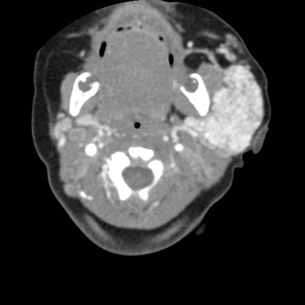
 Cardiac Hemangioma Radiology Case Radiopaedia Org

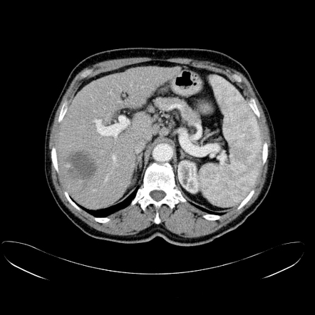
 Hepatic Hemangioma Radiology Reference Article Radiopaedia Org
 Symptomatic Thoracic Vertebral Hemangioma A Case Report And Literature Review1 Archives Of Physical Medicine And Rehabilitation
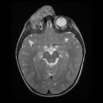
 Orbital Cavernous Haemangioma Radiology Case Radiopaedia Org Radiology Radiography Radiologic Technology
 Infantile Hemangioma Radiology Reference Article Radiopaedia Org
 Hepatic Hemangioma Radiology Reference Article Radiopaedia Org
 Hemangioma Liver Lesions
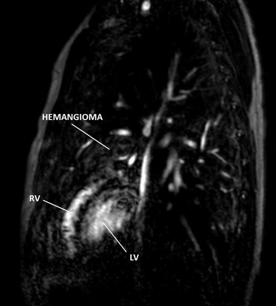 Cardiac Hemangioma Radiology Case Radiopaedia Org
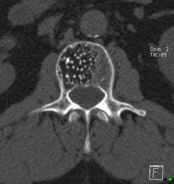 Vertebral Hemangioma Radiology Reference Article Radiopaedia Org
 Atypical Mr Imaging Features Of Vertebral Hemangioma Involving The T12 Download Scientific Diagram
Hemangioma Radiology Reference Article Radiopaedia Org
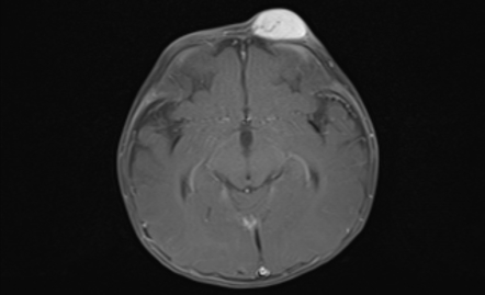 Infantile Hemangioma Radiology Reference Article Radiopaedia Org

 Infantile Hemangioma Radiology Reference Article Radiopaedia Org
 Infantile Hepatic Hemangioma Radiology Reference Article Radiopaedia Org
Https Www Ajronline Org Doi Pdfplus 10 2214 Ajr 180 1 1800135
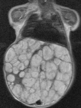
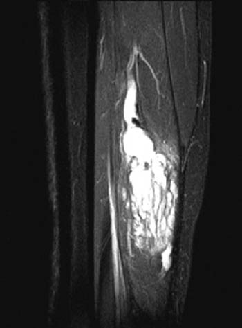 Venous Malformations Radsource
 Internal Hemangiomas Types Diagnosis And Treatment